Powdery Mildew and Fungicides
Part 3: Plant Breeding
Plant Breeding as a Strategy
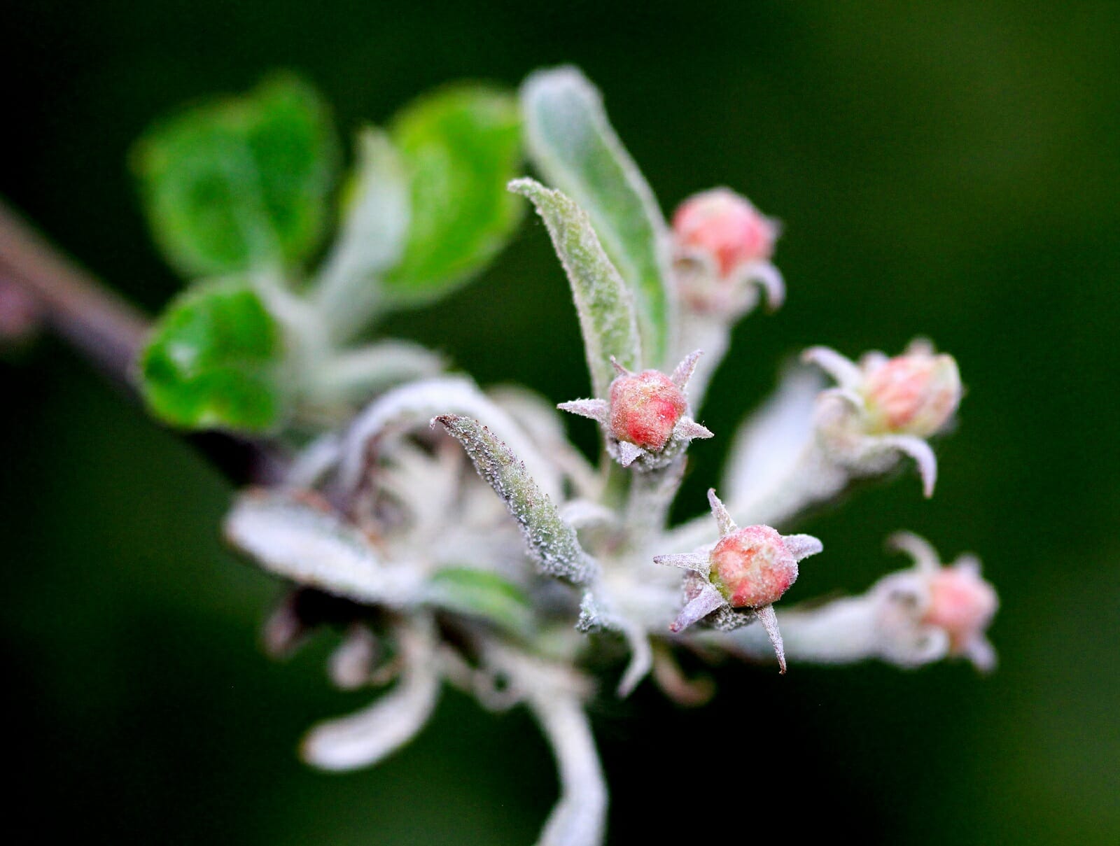
Fungal infection resistance in any particular strain of plant can range from immunity (no infection at all) to resistant (some infection) to susceptible (significant infection). Plants that show definite infection resistance during an epidemic can be removed from the immediate danger and used in one way or another as breeding stock.
A planned breeding strategy is resource-consuming beyond the capacity of most growers. But be aware that some people are doing this. Plants do exist that possess fungus resistant genetics. This often involves changes to the composition of the epidermal cell’s exterior wall. The cell wall is made more resistant to enzymatic breakdown, thereby making it much harder for the developing spore to get its “hooks” into the plant cell. Other resistant plants have developed enzymes that break down fungal cell walls or fungal hyphal tips.
Powdery Mildew Lifecycle (Advanced Level)
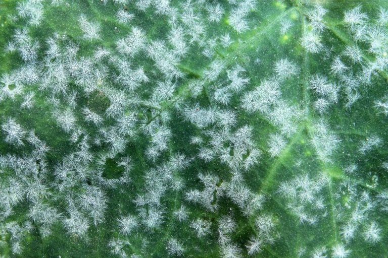
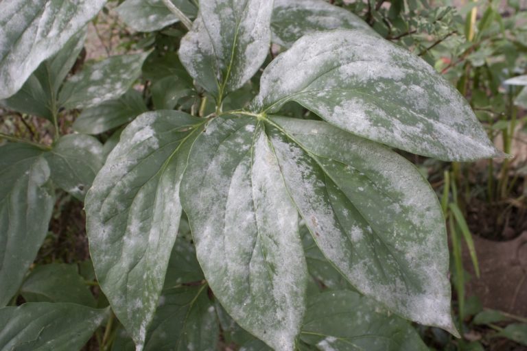
The white “powder” isn’t powder, it’s the mildew’s growth and reproductive system. Keep in mind that mildew is mold, and they are all fungi. Powdery mildew is a fungus.
Your fungal infection—mildew—can start soon after spores brought into your grow room land on leaf surfaces. Powdery mildew spores are well stocked with water and fat content. Therefore, powdery mildew does not require liquid water to germinate. The spores germinate under favorable conditions of 60° to 70°F, dry leaves, and around/over 60%RH. Germination and growth show the small, white, circular patches of fuzz that can be easily wiped off. Don’t be fooled, though… those little patches have already produced spores by the hundreds that are quickly spreading to your other plants.
Fungi break into the plant by using enzymes to soften the epidermal cell walls and mechanical pressure to push through. There they find nutrients supplied by the plant’s vascular system. As the infection progresses, the fungus spreads toward the tops of the plants and into any available buds.
fungal reproductive units
Spores are the fungal reproductive units, they are much like very fast growing seeds. Spores consist of one or a few cells; spores range in size but are often around 0.001” to 0.003” across. Powdery mildew can produce two types of spores—asexual clone spores (conidia), which it produces quickly after establishing itself within the plant; and sexual reproductive spores (ascospores), which it produces when it advances through its later life cycle. Both of these types, clone spores (conidia) and sexually recombined spores (ascospores), have the same goal at their initial germination: grow a powdery mildew individual. The ascospores grow a new genetic individual while the conidia grow a clone of their source individual. Because the genetic recombination of sexual reproduction produces new genotypes, some of these new individuals may be more resistant to fungicides or other eradication practices than either of the parental genotypes.

Life cycle of powdery mildew
Image adapted from: Agrios, G.N. 1997. Plant Pathology. 4th edition.
Hypha are the thread-like filaments formed by a germinating spore (multiple hypha are hyphae), the developing hyphal mat is called a mycelium (the whitish looking mycelium is the first visible indication of the infection). The first type of hyphae produced by germinating spores are called germ tubes, these penetrate the plant’s epidermal cells. A haustorium is the hyphal structure that grows inside a host epidermal cell in order to more effectively steal nutrients. A conidiophore is a specialized hypha that grows vertically and produces the vegetative spores (conidia). The mass of hyphae, the mycelium, comprises the vegetative body of the fungus.

Powdery mildew fungal infection: Cuticle and epidermal cells penetrated.
The plant cell wall is not simply a passive barrier, it is a dynamic structure that can be actively reinforced in local regions under certain types of attack.
immune responses
Plants show 2 types of immune response—pattern triggered immunity (or, basal immunity), which results in local reinforcement of the cell wall (often modifications to pectin); and the aptly-termed hypersensitive response, which kills cells that are under attack. Pattern triggered immunity is triggered when plant cells recognize certain “microbe-associated molecular patterns” (MAMPs) including specific molecules and cell wall components commonly found in microbes. Hypersensitive response can occur as a reaction to infection peg penetration, or haustorial growth; it is a very effective defense in capable plants.
hyphae
Hyphae are capable of developing a variety of forms under genetic control. Fungi can and do alter the developmental pathway of individual hyphae in response to environmental changes. For instance, the growing fungal germ tube perceives physical cues from the leaf surface. These cues determine when the tip of the germ tube stops growing outward. As outward growth ceases the germ tube hooks toward the leaf and begins to swell. The germ tube tip cells go through a differentiation process forming a structure called an appressorium. An appressorium is a highly specialized infection cell. Substances accumulate within the appressorium which increase its turgor pressure. This pressure increase inside the appressorium works with cell wall dissolving enzymes to help to drive what’s sometimes known as an “infection peg” through the plant cuticle and into the plant’s epidermal cells.

One example of a plant’s basal immunity: Callose deposits.
As an example of pattern triggered immunity, plant epidermal cells synthesize and deposit callose which form papillae (much like our own callouses) to fight the attacking mold. If the flat, thick papillae are compromised, the plant can form haustorial collars and haustorial encasements as another defensive response to hyphal penetration. Haustorial collars are nooses around the haustorial feeding tube, while haustorial encasements are the plant’s attempt to put a bag around the haustorium. These defensive structures can be chemically suppressed by some fungi. There is constant life and death battle between plant and fungus.


Examples of plant defenses against fungal infection.
conidia
Conidia (genetically identical to the individual mycelium from which they originate) are usually produced within a few days of successful spore germination. They are colorless, and generally oval in shape. The exposed hyphae grow many conidiophores that produce conidia either singularly or chained, depending on the species of fungus. They are produced as long as (and whenever) environmental conditions are favorable. Conidia excrete an adhesive soon after landing on a leaf surface, this adhesive facilitates recognition of a host plant, which cues subsequent germination. Conidia production is part of the asexual development stage of the powdery mildew fungus.


Asexually produced spores (clones of the individual)
Outdoors, as the season changes, the fungus moves into its sexual reproductive stage. This stage is rarely, if ever, seen (allowed to occur) indoors.
Production of conidia decreases and eventually halts going into fall as the weather turns cooler. Powdery mildew fungi then produce relatively large reproductive (fruiting) bodies called ascocarps. Ascocarps function to hold a type of special container cell called an ascus (plural: asci). Asci contain the sexually produced spores, the ascospores. There are four different configurations of ascocarps. The generally-spherical ascocarp produced by powdery mildew is called a cleistothecium (clice-toe-th-ee-see-um). Cleistothecia are only formed during the sexual reproductive phase of the powdery mildew lifecycle. Ascocarps also allow the fungus to hibernate through poor conditions as they are very resistant to low temperatures and drought.
reproduction
Sexual reproduction begins when a receptor on one haploid hypha detects a pheromone from a complementary mating type. Haploid means it has its usual single set of fungal chromosomes. Specific pathways are activated inside the hyphal cells, and the hypha approaches the source of the pheromone through chemotropic growth. The fungus then exposes it’s mating type by developing one of two complementary organs, a “female” (+) ascogonium or a “male” (-) antheridium.
These reproductive organs contain only haploid (single set of fungal chromosomes) cell nuclei. A bridge (the trichogyne) forms between them, providing a path for nuclei to move from the antheridium to the ascogonium. A dikaryote (has 2 nuclei) then grows from the ascogonium. Karyogamy (fusion of the 2 haploid nuclei) then occurs, after which the diploid (has 2 sets of chromosomes that are joined) zygote undergoes meiotic divisions to yield the haploid ascospores. The ascospores go through a mitotic (simple clonal growth) division that double their number. All this activity during and after sexual recombination occurs within the newly formed cleistothecium.
As an interesting note: Depending upon the species of Erysiphaceae, the fungus can be homothallic or heterothallic. Heterothallic means the fungus require two compatible partners to perform sexual reproduction. A homothallic fungus is capable of sexual reproduction all by itself. Homothallism has less genetic recombination and may be more of a repair process.

– (antheridium) and + (ascogonium) reproductive organs showing trichogyne (nuclear transfer bridge) and dikaryotic hyphae
chromosome path

Chromosome path for sexual reproduction: From 2 mating individuals to 4 new genetic individuals
Description of the “Chromosome path” diagram, above: The set of chromosomes contributed by the male merges with those contributed by the female in what is termed a nuclear union (because chromosomes are located in the nucleus of the cells). During the interphase stage of meiosis, the chromosomes duplicate and the homologous chromosomes (m/f chromosomes of the same type) exchange genetic information (chromosomal gene crossover) during the first division, called meiosis I. The daughter cells divide again in meiosis II, splitting up the newly formed “sister chromatids” in order to return once again to the usual haploid cells.
These cells go through a cloning (mitotic) reproduction within the ascus structure to double their number from 4 to 8 (this is depicted in the diagram below). They then mature into ascospores, ready for the cleistothecium to break open and their asci to release them into the breeze.

Generic ascocarp showing complete sequence of ascospores production.
cleistothecia
Cleistothecia first appear white, turning yellow, orange, brown, and eventually black as they mature in/on the mycelial mats. They develop throughout the fall and are mature over winter. When conditions are favorable, the cleistothecia absorb water and burst open to expel their contents (the asci, or container cells). The asci burst open and discharge the ascospores, which become windborne to eventually germinate on a host plant.


Immature and mature cleistothecia on leaf
When conidia or ascospores contact a suitable host’s leaf (top or bottom) they will germinate within 48 hours if temperature and RH are sufficient. Each spore produces a germ tube that eventually generates a short, thin hypha that attempts to pierce the host cell cuticle, and epidermal cell wall, to enter an epidermal cell. On entry into an epidermal cell, the hypha forms the feeding organ, the haustorium. Haustoria rob nutrients from the plant’s epidermal cells; nutrients that you and your plant have worked so hard to obtain.

Another PM lifecycle diagram: Exercise your new knowledge with this version.
Powdery Mildew Taxonomy
| Kingdom: | Fungi |
| Phylum: | Ascomycota |
| Class: | Leotiomycetes |
| Order: | Erysiphales |
| Family: | Erysiphaceae |
| Genus: | ~28 genera
Examples- Brasiliomyces, Podosphaera, Sphaerotheca, Uncinula, Phyllactinia, Oidium |
| Species: | ~700 species |
Note: All pictures and diagrams here were originally produced by others. They are the clearest and best examples available with which to educate and inform. Some were slightly adapted for this use.
This educational document was written, compiled, and adapted by Mike Steffes of Quest Dehumidifiers.
Powdery Mildew and Fungicides Parts 1–3

Explore how plant breeding serves as a long-term strategy for combating powdery mildew. By selecting for genetic resistance, growers can develop plants with reinforced cell walls that deter fungal penetration. This post also dives into the advanced lifecycle of powdery mildew, examining how spores germinate, spread, and evolve to survive your best eradication efforts.
Managing powdery mildew requires effective treatment options ranging from inorganic chemicals to organic solutions. Explore the benefits of potassium bicarbonate, sulfur vaporization, and various oils like neem or coriander. Whether using hydrogen peroxide or milk dilutions, understanding the specific mechanisms behind these fungicides can help you protect your plants from persistent fungal infections efficiently.
Powdery mildew is a common fungal disease caused by the Erysiphaceae family. As an ectoparasite, this fungus grows on the surface rather than inside plant tissue. While not systemic, it spreads rapidly through spores, stunted growth, and high humidity. Learn the fundamentals of identifying, managing, and preventing this persistent plant pathogen in your garden.
Published on Jun 26 2017
Last Updated on Jun 05 2026
Categories: Agriculture, Bud Rot, Cannabis Mold, Mildew, Mold, Powdery Mildew